Ultrasound imaging is one of the most widely used diagnostic tools in modern medicine, primarily because it is safe, painless, non-invasive, and highly effective for examining abdominal and pelvic organs. However, what many patients—and sometimes even clinical staff—tend to overlook is the importance of timing. When an ultrasound is performed at the right moment, the images become significantly clearer, more detailed, and easier to interpret. Small physiological changes—like the presence of bowel gas, bladder fullness, or hormonal variations throughout the menstrual cycle—can dramatically impact visibility.
Understanding when to schedule an abdominal or pelvic ultrasound helps clinicians reduce artifacts, shorten scanning time, lower the likelihood of repeat exams, and increase diagnostic accuracy. This article explains the best timing strategies for both abdominal and pelvic imaging and highlights how certain clinical factors influence scheduling decisions.
Why Timing Influences Ultrasound Quality
The effectiveness of ultrasound depends on how easily sound waves can travel through body tissues. When gas, empty organs, or fluctuating hormone levels interfere with this pathway, the resulting images may be unclear or incomplete.
For abdominal exams, excessive bowel gas is one of the most common obstacles. Gas blocks sound waves, preventing the sonographer from obtaining clear views of the liver, pancreas, gallbladder, and bile ducts. Similarly, food intake triggers digestive activity, making organs move and contract—further complicating imaging.
Pelvic imaging is influenced by additional factors, such as hydration and hormonal cycles. A properly filled bladder acts as an acoustic window, pushing bowel loops away so the ultrasound beam can reach the uterus and ovaries. On the other hand, the menstrual cycle causes the endometrium and ovaries to change shape and thickness, affecting how well they can be seen.
All these elements demonstrate why choosing the right moment is essential for obtaining high-quality diagnostic results.
The Best Time for an Abdominal Ultrasound
When scheduling an abdominal ultrasound, timing and preparation go hand in hand. The general recommendation is to perform the scan early in the morning. This aligns with a natural period of fasting, which prevents bowel gas from accumulating and helps the gallbladder remain fully distended.
A fasting period of 6–8 hours is considered ideal. This allows the stomach and intestines to empty and reduces the likelihood of gas interference. It also ensures the gallbladder is filled with bile, providing a much clearer view of its walls and contents.
Patients undergoing evaluation for gallstones, fatty liver disease, bile duct obstruction, or pancreatic issues benefit significantly from morning scans.
Despite these guidelines, emergencies must always override timing rules. For example:
- Severe abdominal pain
- Suspected appendicitis
- Internal bleeding
- Signs of acute gallbladder inflammation
In these scenarios, immediate imaging is prioritized because the patient’s stability is more important than ideal visualization conditions. Sonographers adjust their scanning techniques to optimize images even when timing cannot be controlled.
The Best Time for a Pelvic Ultrasound
Pelvic ultrasounds, used to assess the uterus, ovaries, bladder, and surrounding structures, require a different approach. The optimal timing depends on the type of pelvic exam and the reason for the evaluation.
Transabdominal Pelvic Ultrasound
For this method, the patient must have a full bladder. A filled bladder acts like a natural lens, improving the transmission of sound waves and gently displacing bowel loops.
Patients are typically instructed to drink 4–6 cups of water about one hour before the scan and avoid urinating until the exam is complete. This simple preparation results in dramatically improved image clarity.
Transvaginal Pelvic Ultrasound
Bladder filling is not needed for this method, but menstrual cycle timing matters:
- Early to mid cycle (days 5–14): Best for evaluating ovarian follicles, ovulation patterns, or follicular growth.
- Late cycle: Best for assessing endometrial thickness, implantation conditions, and hormone-related changes.
- Any time symptoms arise: For pain, abnormal bleeding, or suspected cysts, the scan is performed based on clinical need rather than cycle timing.
Because reproductive organs undergo dynamic changes throughout the month, scheduling the scan correctly allows clinicians to obtain the most accurate representation of pelvic anatomy.
Key Factors That Modify the Recommended Timing
Although general guidelines exist, patient-specific factors often influence when an ultrasound should be performed. These include:
Medical History
Patients with chronic gallbladder disease, PCOS, uterine fibroids, or hormonal disorders may need scheduled imaging at particular phases of the cycle or after specific periods of rest or fasting.
Pregnancy and Reproductive Status
Pelvic scans differ significantly between premenopausal and postmenopausal patients.
- Pregnant patients may require imaging tied to fetal development stages.
- Postmenopausal patients often have more consistent anatomy, making timing less restrictive.
Symptom Severity
If symptoms are mild and stable, clinicians follow ideal timing guidelines.
If symptoms are severe, the scan becomes urgent regardless of preparation.
Suspected Diagnosis
Some conditions—such as ovarian torsion, ectopic pregnancy, and biliary obstruction—require immediate imaging.
Clinical Urgency
When clinicians suspect life-threatening issues, they prioritize rapid intervention over timing optimization.
Modern Ultrasound Devices and Their Effect on Timing
Today’s imaging technology offers more flexibility in when and where ultrasound exams can be performed. While timing remains important for maximizing clarity, device choice also plays a role—especially in fast-paced medical settings.
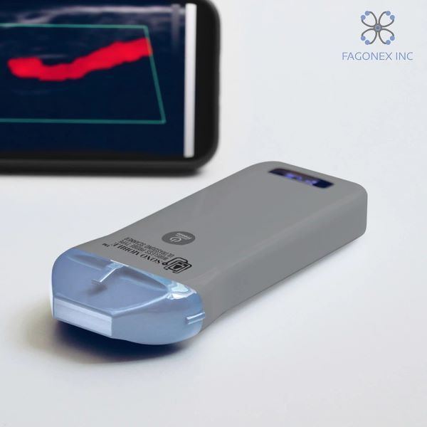
In emergency departments, urgent-care clinics, and bedside assessments, healthcare professionals increasingly rely on handheld ultrasound devices. These compact systems enable rapid, point-of-care evaluation without waiting for the patient to reach a dedicated imaging room.
Laptop-sized portable systems offer stronger processing power and deeper resolution, making them ideal for outpatient clinics and routine diagnostics. Traditional cart-based ultrasound machines remain the gold standard for high-resolution imaging, especially when deep structures or subtle abnormalities must be evaluated.
Although newer tools improve accessibility and speed, timing principles—such as fasting or cycle scheduling—still produce the clearest results when conditions allow.
When You Shouldn’t Delay an Ultrasound
Certain clinical scenarios override timing recommendations due to the risk of complications. Immediate imaging is necessary in cases such as:
- Severe abdominal or pelvic pain
- Heavy vaginal bleeding
- Suspected ectopic pregnancy
- High fever with localized tenderness
- Potential internal bleeding
- Sudden right upper quadrant pain
- Acute urinary obstruction
In these situations, clinicians perform the scan immediately, adjusting scanning angles and techniques to reduce artifacts even without ideal conditions.
Conclusion
Scheduling an abdominal or pelvic ultrasound at the right time can dramatically improve image accuracy and clinical interpretation. Whether it’s fasting for an abdominal scan or ensuring proper hydration for a pelvic evaluation, small adjustments in timing create measurable improvements in diagnostic clarity. Modern ultrasound technologies offer new levels of convenience, but anatomical and physiological patterns still guide the best moments for imaging. By understanding these principles, both clinicians and patients can support better decision-making and minimize the need for repeated exams, ultimately leading to more effective and timely care.


پاسخ ها